Treatment of the temple with hyaluronic acid filler: penetration force through the superficial and deep temporal fascia and implications for treatment
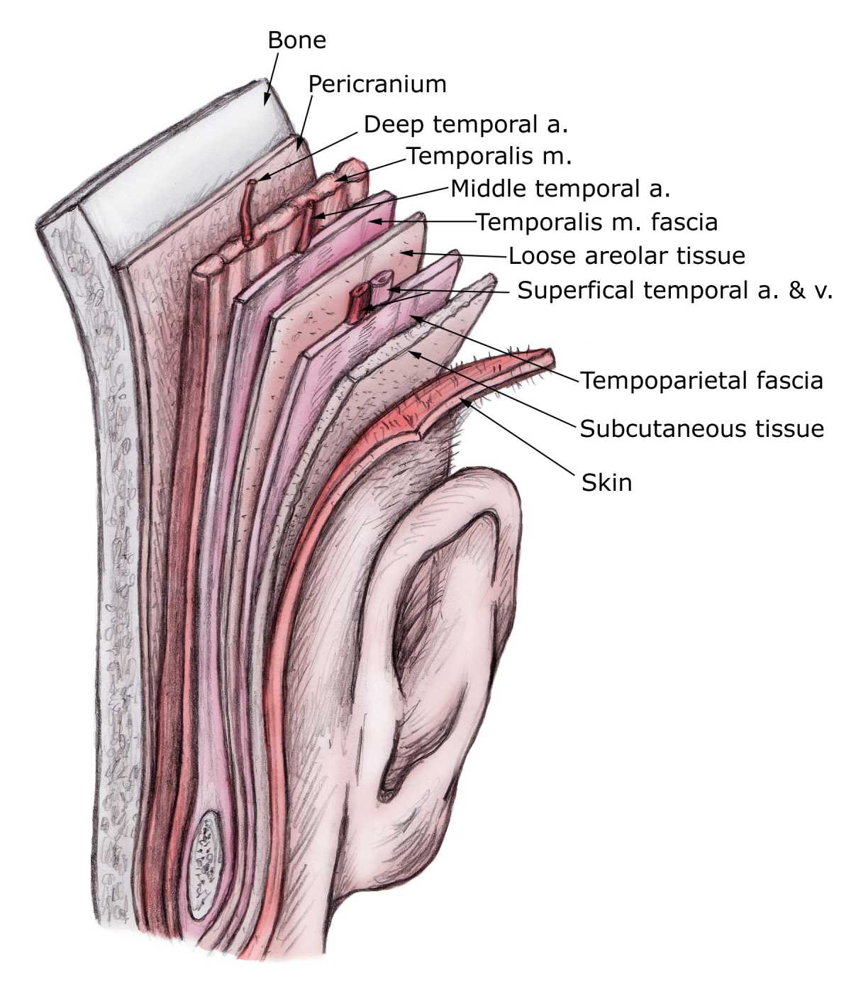
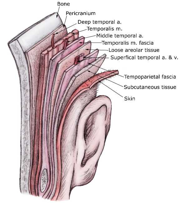
Figure 1: The temporoparietal fascial flap.
Volumisation of the temple region is one of the most frequently performed treatments with hyaluronic acid fillers. However, a wide variety of techniques with HA fillers are recommended in the literature. Some studies, for example, propose injecting the filler deep into the periosteal layer, between the temporal fossa and the temporalis muscle, in the depressed area. However, this technique requires a relatively large amount of filler material, and it is not always possible to fill the entire depression effectively and without complications.
An alternative and frequently performed technique is injection of the filler material between the superficial and deep temporal fascia (Fig. 2).
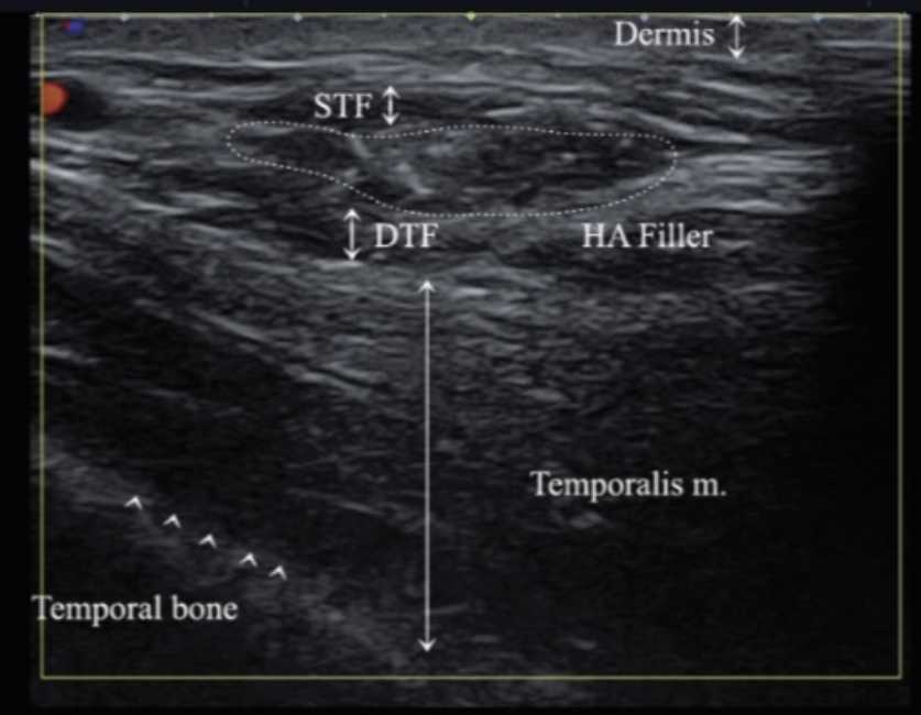
Figure 2: Ultrasound image of a hyaluronic acid filler (HA filler) injected between the superficial temporal fascia (STF) and the deep temporal fascia (DTF).
For effective and safe treatment with this technique, however, a precise understanding of the layered structure of the temple area is essential. Additional quantitative information can increase the efficacy and safety of this technique. A recently published study (Lee et al. J Plast Reconstr Aesthet Surg . 2022 Dec 19;77:271-273), for example, examined the penetration force needed to penetrate the superficial and deep temporal fascia. Conclusions for treatment were drawn from the results.
Materials and methods:
The superficial and the deep temporal fascia were exposed by removing the skin and the subcutaneous tissue around the temple area, and each was retracted. The penetration forces required to penetrate the two fasciae were measured (Fig. 3).
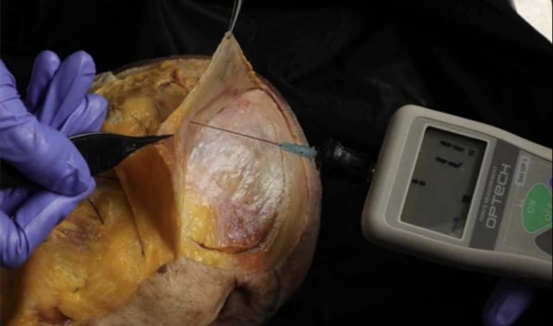
The upper point was set 2 cm lateral to the lateral eyebrow and the lower point 2 cm above the zygomatic arch on a line perpendicular to the upper point. The penetration force was measured with a 21G cannula on a multiTest 2.5-d device (Mecmesin Limited). To avoid potentially confounding factors, the fascia under examination was firmly fixed throughout the entire process. The mean value was determined by measuring the left and right sides three times.
Results:
At the upper point, the mean penetration force was 7.5 ± 2.9 N for the superficial temporal fascia and 13.2 ± 3.2 N for the deep temporal fascia. The mean penetration force at the lower point of the deep temporal fascia was 11.5 ± 3.3 N.
Discussion:
The present study shows that it is not easy to penetrate the deep temporal fascia.
As the penetration force through the superficial fascia differs from the penetration force through the deep temporal fascia, this fact can serve as an aid for practitioners in distinguishing the two structures from each other by feel.
Placing the cannula tip between the two fasciae is actually a relatively safe technique. However, the middle temporal vein is known to lie deep in the superficial layer of the deep temporal fascia, so that there is a risk of vascular injury with deep injections. It can therefore be useful to locate vessels in the temple area with Doppler ultrasound.
In addition, it may be safer to use a cannula with a larger diameter. For example, it is likely that the patient will feel the perforation of the superficial temporal fascia when a 21G cannula is used.
Source: Lee et al. J Plast Reconstr Aesthet Surg . 2022 Dec 19;77:271-273